Take-home points
|
 |
Bios Dr. Singaravelu is a vitreoretinal surgery fellow with the Retina Group of Washington and MedStar Georgetown University Hospital, Washington, D.C. Dr. Do is a vitreoretinal surgeon and uveitis specialist at the Retina Group of Washington and is a clinical assistant professor at Georgetown University School of Medicine. He is also a member of the executive committee of the Vit Buckle Society. DISCLOSURES: Dr. Do disclosed financial relationships with AbbVie/Allergan, Alimera Sciences, Bausch + Lomb Pharmaceuticals and Bausch + Lomb Surgical. |
The 12th Annual Meeting of the Vit Buckle Society, held in Miami, highlighted vigorous and animated surgical discussion among both trainees and post-training vitreoretinal surgeons from all over the world. Here, we present pearls for dealing with vitreoretinal surgical challenges from 14 of them.
These pearls include a novel approach to lensectomy, tips for surgery for myopic traction maculopathy, retinal detachment after globe rupture and intraocular foreign body removal, as well as managing a host of unique presentations—chronic detachment with macrocysts, subfoveal subretinal bands and metastatic cutaneous and ciliary body melanoma.
A separate session featured vitreoretinal surgeons from Mexico and Canada sharing their approaches to seven surgical dilemmas: internal limiting membrane flap complications; diabetic tractional retinal detachments; rhegmatogenous retinal detachments; implants for macular buckling; endoscopic visualization for anterior proliferation; addressing the anterior hyaloid in pediatric vitrectomy; and creative ways of managing macular holes.
 |
Novel strategies for removing dense subluxed cataracts
Samir Patel, MD, from Retina Vitreous Consultants in Pittsburgh, discussed a novel approach to lensectomy. He presented a case of a monocular patient with a history of pathologic myopia and pseudoexfoliation glaucoma with a dislocated crystalline lens that was too dense to be removed with a fragmatome.
After performing a vitrectomy, Dr. Patel explained how he used an intracapsular approach with the assistance of perfluoron to levitate the lens just below the iris plane. He then used a lens loop to elegantly remove the entire cataract through a scleral tunnel.
Mrinali Gupta, MD, of Retina Associates of Orange County in Southern California, shared her experience with a similar case in which she used a cryotherapy probe to engage the lens and remove it through a scleral wound.
 |
Macular surgery in myopic traction maculopathy
Vitreoretinal surgery in myopic eyes can be challenging. Seasoned vitreoretinal surgeons shared their pearls and some words of wisdom for managing these cases.
Cassie Ludwig, MD, of the Byers Eye Institute at the Stanford University School of Medicine in Palo Alto, California, shared her expertise on surgery for myopic traction maculopathy with a fovea-sparing internal limiting membrane peel for foveoschisis retinal detachment. She underscored the importance of carefully lifting the posterior hyaloid due to its taut nature, restaining with triamcinolone to identify vitreoschisis, and assessing whether an iatrogenic macular hole has been created.
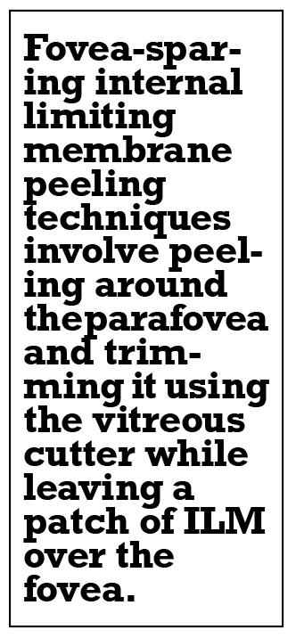 |
Fovea-sparing ILM peel techniques involve peeling around the parafovea and trimming it using the vitreous cutter while leaving a patch of ILM over the fovea, Dr. Ludwig said. An advantage of this technique is that it avoids macular hole formation, but a disadvantage is its potential to enable formation of an epiretinal membrane over the fovea. She used long-acting C3F8 gas with extended positioning to achieve a successful outcome.
Session comoderator Carl Regillo, MD, chief of the retina service at Wills Eye Hospital/Mid Atlantic Retina in Philadelphia, offered these additional pearls for vitreoretinal surgery in myopic eyes with long axial length: using 23-gauge instrumentation along with extra-long myopic instruments; and decreasing the infusion pressure to soften the globe and allow for greater indentation.
Yoshi Yonekawa, MD, an adult and pediatric retina specialist at Wills Eye/Mid Atlantic Retina, noted that while foveal ERM formation is always a risk, it’s typically mild and doesn’t require any further surgery.
 |
Tips for traumatic retinal detachments in repaired ruptured globes
Kirk Hou, MD, PhD, of Doheny Eye Institute UCLA in Los Angeles, presented a case of traumatic retinal detachment in a patient with a history of a ruptured globe with a repaired corneal laceration, resulting in a limited view to the posterior pole.
After he removed the residual lens material and vitreous hemorrhage, he noted a total retinal detachment with a star-fold, retinal foreshortening, along with an area of chorioretinal incarceration at the site of a posterior perforation. He used perfluoron and performed an inferior 180-degree retinectomy and focal retinectomy incorporating the area of incarceration. Silicone oil provided a tamponade agent, after which the retina remained attached.
Jayanth Sridhar, MD, an associate professor of clinical ophthalmology at Bascom Palmer Eye Institute in Miami, provided an update that demonstrated the retina remaining attached four months after surgery, and indicated the patient is currently awaiting a corneal transplant.
 |
Removing a metallic intraocular foreign body from the inferior retina
Lediana Goduni, MD, of Retina Associates of Cleveland, shared pearls for removing a metallic intraocular foreign body from the inferior retina. After a thorough vitrectomy, she applied endolaser pulses around the IOFB impact site. Next, she removed the superotemporal cannula and enlarged the sclerotomy. She then removed the IOFB using Maxgrip forceps. C3F8 gas provided an endotamponade and the patient had intravitreal antibiotics.
Dr. Goduni offered this tip: Use computed tomography to estimate the size of the IOFB, which will help to approximate the size of the sclerotomy needed to remove the IOFB during surgery.
Dr. Regillo emphasized the importance of a circumferential pars plana scleral incision for IOFB removal when the wound requires extension. Shilpa Desai, MD, the session’s comoderator and assistant professor at Tufts University School of Medicine in Boston, said an intraocular magnet could be an alternative method for IOFB removal.
 |
Managing a chronic traumatic retinal detachment with macrocysts
Phoebe L. Mellen, MD, of Retina Vitreous Consultants in Pittsburgh, presented the case of a young patient with a chronic traumatic retinal detachment with two macrocysts. The initial repair with primary buckle and gas failed, so Dr. Mellen pursued a vitrectomy, which she completed with the assistance of triamcinolone.
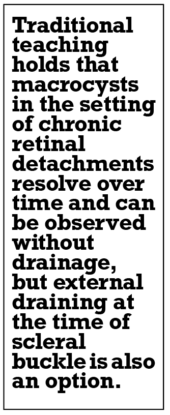 |
Next, Dr. Mellen used diathermy to drain the posterior macrocyst and create a drainage retinotomy to flatten the retina. After air-fluid exchange and endolaser, one large cyst remained, but she was concerned it would tent the retina, preventing adequate closure of the associated breaks. So, she drained the cyst and instilled silicone oil. The retina remained attached two months after surgery without cyst recurrence.
Dr. Regillo and Harry Flynn, MD, professor of ophthalmology at Bascom Palmer Eye Institute, noted that traditional teaching holds that macrocysts in the setting of chronic retinal detachments resolve over time and can be observed without drainage. Dr. Flynn also recommended external drainage at the time of primary scleral buckle.
 |
Managing recurrent RD with subretinal bands
Joshua H. Uhr, MD, of Retinal and Ophthalmic Consultants in New Jersey, discussed a case of recurrent retinal detachment with extensive submacular PVR in the form of subretinal bands. Because of concern that the subretinal bands would impede foveal reattachment, he created retinotomies and used Maxgrip forceps with gentle countertraction from the light pipe to remove the subretinal bands.
 |
Dr. Desai recommended a “spaghetti” technique, using a twirling motion with the forceps while removing long segments of subretinal bands.
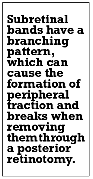
Hong-Uyen Hua, MD, a pediatric retina and vitreous disease specialist at Bascom Palmer, cautioned that subretinal bands have a branching pattern, which can cause the formation of peripheral traction and breaks when removing them through a posterior retinotomy.
 |
Metastatic disease masquerading as uveitis
Jordan D. Deaner, MD, of Wills Eye/Mid Atlantic Retina, presented a case of a patient with metastatic cutaneous and ciliary body melanoma previously treated with excision of the cutaneous lesion, plaque brachytherapy and adjuvant immunotherapy, and who had worsening floaters.
He described atypical vitritis with “large clumps of vitreous cells and debris.” Ancillary imaging studies weren’t consistent with ocular inflammatory disease, so he performed a diagnostic vitrectomy.
The technique involved a thorough vitrectomy using a 23-ga instrumentation at a low cut-rate, collection of undiluted and diluted samples, partial air-fluid exchange with suturing of sclerotomies, and double-freeze thaw cryotherapy to prevent seeding.
Pathology revealed isolated vitreous metastates of cutaneous melanoma. The patient was treated with a series of five monthly intravitreal melphalan injections. The tumor burden resolved and the visual outcome was excellent.
Basil K. Williams Jr., MD, associate professor at Bascom Palmer, offered a diagnostic clue to differentiate metastatic cutaneous melanoma vitreous seeding from uveitis: the pigmented appearance of vitreous cells. RS
Surgical pearls from north and south of the border By Jovi Wong, MD, MSc, PhD; Aditya Rali, MD, Luke Oh, MD, and Brian K. Do, MD
In Mexico and Canada they do things a little differently from the United States, especially when it comes to retinal surgery. Vitreoretinal surgeons from across both borders shared their approaches to a host of challenging surgical scenarios, providing a study in contrasts in techniques. Avoiding and dealing with ILM flap complications Virgilio Morales-Canton, MD, of the Association to Prevent Blindness Mexico, demonstrated how he dealt with a displaced internal limiting membrane free flap during repair of a large macular hole in a myopic adult patient. He discussed a case in which the flap became displaced during air-fluid exchange. The patient had a postoperative retinal detachment secondary to the large macular hole. In a subsequent operation, Dr. Morales-Canton performed the air-fluid exchange through the macular hole, then lasered the macular hole edges to secure the retina and repair the detachment. The patient ended up with 20/50 visual acuity. In cases of lost ILM flaps, Dr. Morales-Canton offered these options for macular hole closure: hinged ILM flaps; ILM free flaps; amniotic membrane grafts; and autologous retinal transplant.
Parnian Arjmand, MD, of the Toronto Retina Institute, reviewed a variety of ILM flap complications and tips on how to avoid them. They included avoiding flap amputation, ensuring appropriate—that is, not undersized—ILM flaps, and avoiding flap movement under air. Perioperative solutions she suggested included cutting on zero suction, slowing hand movements with forceps, and leaving multiple flaps available as backups. For her ILM flaps, Dr. Arjmand said she likes to create multiple flaps, alternating clockwise and counterclockwise and creating progressively larger concentric circles. She also demonstrated and advocated for the use of a Tano scraper with viscoelastic to mobilize the ILM flap into position. She noted that patient selection is important. In these cases, she said, Brilliant Blue dye might be preferred over indocyanine green given the higher likelihood of toxicity from the latter. Managing diabetic tractional RDs Gerardo Garcia-Aguirre, MD, of the Institute of Technology in Monterrey, focused on maneuvers for successful diabetic tractional retinal detachment repair. Among 11 suggested plays, his tips included: using a chandelier for every case; pulling up on the tractional membrane at the nerve to look for a plane if one isn’t obviously present (space-maker); reducing the cut rate to help cut through spongy membranes (slow cutter); and using forceps along with the cutter to help hold up a membrane and simultaneously cut through (forceps dissection). The 27-ga cutters are useful in approaching TRDs, Dr. Garcia-Aguirre said, because the shorter length between the end of the vitrectomy probe and cutter opening vs. the 25-ga instruments enables entrance into tighter spaces between the membranes and retina. Dr. Garcia-Aguirre also suggested these two techniques for trimming the isolated fronds of fibrovascular proliferation as closely as possible:
He also emphasized the importance of promptly controlling bleeding by applying diathermy.
A novel approach to RRD repair In-office suprachoroidal viscopexy is a novel technique for rhegmatogenous retinal detachment. Rajeev Muni, MD, of St. Michael’s Hospital in Toronto recommended this technique to reduce unwanted retinal displacement and distortion following conventional RD repair with vitrectomy or scleral buckle. He noted that anatomic reattachment doesn’t necessarily equal success. That’s because commonly used surgical repair techniques involve rapid retinal reattachment and resolution of subretinal fluid, which can lead to unwanted folds and retinal displacement. The postoperative metamorphopsias and aniseikonia that some patients can experience are still anatomically successful but less than desirable functionally, Dr. Muni said. A slow, natural reattachment of the retina ideally shouldn’t require patient positioning or invasive surgery, Dr. Muni said. In the initial case series he presented, injecting viscoelastic into the suprachoroidal space under the retinal break achieved retinal reattachment without complications. Patients with relatively shallow detachments, with breaks outside of the 12 and 6 o’clock positions, are good candidates for this procedure, Dr. Muni said. A different and less invasive approach to macular buckling A new and potentially less invasive option for myopic traction maculopathy are polytetrafluoroethylene (PTFE) felt implants. Gabriela Lopez-Carasa, MD, of Macula Retina Consultants in Huizquilucan, Mexico, explained that these implants, fabricated from material similar to that used in vascular and orbital surgery, are cut to size to suit the patient. The implants are inert, nonantigenic and easily moldable. An accompanying video showed how her technique for placing the implant. She folded a 6 x 12-mm piece of PTFE felt in half, then passed a Mersilene suture through the halves to avoid losing it in the orbit. Dr. Lopez-Carasa used a Snellen loop to push the implant through the sub-Tenon’s space until an indentation appeared under the macula. A postoperative B-scan showed the axial length was shortened from 36 to 32 mm and successful treatment of a macular detachment due to staphyloma. The patient’s visual acuity improved and subretinal fluid resolved after the operation. Endoscopic visualization for anterior proliferation Flavio Rezende, MD, of the University of Montreal, narrated a complicated repair of redetachment under oil with hypotony in a phakic young high myope. After removing the oil, he used bimanual dissection to remove the very sticky hyaloid face and attempted to use perfluorocarbon to help with this dissection, but ultimately had to remove it due to concerns the PFC would migrate subretinally. He did ILM peeling using Brilliant Blue for visualization. Ultimately, he had to use an endoscope to complete the dissection. Under endoscopic visualization, an anterior ring of proliferation along the ciliary body was noted and removed. Dr. Rezende said these membranes were thought to have contributed to the patient’s preoperative hypotony and likely would not have been addressed using traditional “top-down” visualization. Notably, although iatrogenic retinal breaks were sustained, Dr. Rezende didn’t use endolaser in a move to avoid further retinal contraction. Ultimately, oil was put back into the eye, postoperative methotrexate injections were employed, and the patient’s retina was ultimately noted to have successfully reattached, with a final visual acuity of 20/300 under silicone oil. The importance of addressing the anterior hyaloid When performing pediatric vitrectomy, leave the posterior hyaloid alone, Maria Martinez-Castellanos, MD, of the Association to Prevent Blindness in Mexico, advised attendees. Doing so will avoid inadvertent iatrogenic breaks, proinflammatory bleeding, and the need for retinal laser, she said. The anterior hyaloid, on the other hand, is the most important part of the eye to address in familial exudative retinopathy, retinopathy of prematurity or after retina or congenital cataract surgery. Dr. Martinez-Castellanos noted that these membranes must be removed meticulously to reduce abnormal traction, inflammation and development of fibrovascular tissue. In cases in which the anterior hyaloid is adherent to the posterior lens capsule, removal of the crystalline lens might be necessary, she said.
'Plugs' as autografts to creatively manage macular holes Efrem Mandelcorn, MD, associate professor at the University of Toronto, presented two cases of macular holes and the unique “plugs” he has used as autografts. The first case involved development of an inadvertent iatrogenic macular hole during TRD repair. He placed in the macular hole fibrovascular tissue that had been dissected during the repair. The hole closed postoperatively. The second case involved a patient who needed multiple secondary surgeries after primary pars plana vitrectomy for a giant retinal tear-associated RD. The patient developed two macular holes under silicone oil and underwent an amniotic membrane transplant. However, the eye developed an inferior RD, thought to be secondary to contraction of the amniotic membrane and development of an inferior break along the inferior edge. As a result, the macular holes didn’t fully close. Dr. Mandelcorn employed two peripheral retinal autografts, and the retina ultimately remained reattached, even after silicone oil removal. Final visual acuity was 20/200. BiosDr. Wong is a resident physician in the department of ophthalmology and vision sciences at the University of Toronto. Dr. Rali is a vitreoretinal surgery fellow at the Retina Group of Washington MedStar Georgetown University Hospital in Washington, D.C. Dr. Oh is an ophthalmology resident at Columbia University Irving Medical Center in New York. DISCLOSURES: Dr. Wong, Dr. Rali and Dr. Oh have no financial disclosures. |