Take-home points
|
 |
Bios Mr. Coovadia is an associate of Manhattan Retina and Eye Consultants, New York, and a graduate student at Loyola University Chicago. Ms. Shah is a medical student at Stanford University School of Medicine, Palo Alto, California. Dr. Parikh is chairman of Manhattan Retina and Eye Consultants and director of health-care delivery research in the department of ophthalmology at New York University Grossman School of Medicine. DISCLOSURES: Mr. Coovadia and Ms. Shah have no relevant disclosures. Dr. Parikh disclosed financial relationships with Anthem Blue Cross and Blue Shield, Apellis Pharmaceuticals, Regeneron Pharmaceuticals, GLG, Health and Wellness Partners, and Axon Advisors, and receives funding from the American Academy of Ophthalmology for work with the Relative Value Update Committee. |
In the 1950s, one of the first uses of telemedicine happened at the Nebraska Psychiatric Institute and Norfolk State Hospital where patients received psychiatric evaluations through closed circuit television.1
Technology has since advanced a tremendous amount, opening up new methods of communication and consequently new ways of patient-doctor interaction. With the smart phone and applications such as Skype, FaceTime and Zoom, physicians have become much more accessible to patients.
But has this accessibility been taken advantage of? And what do these new forms of communication mean for the remote practice of ophthalmology? Up until the recent COVID-19 pandemic, video calls weren’t often used in synchronous visits, with most physicians opting for communication via telephone. However, after the public health emergency, the telehealth landscape changed considerably.2
Here, we consider the current state of artificial intelligence utilization and teleophthalmology across two important retinal pathologies: diabetic retinopathy and age-related macular degeneration. Although both diseases have reliable and effective treatments, many patients, for a host of reasons, don’t get adequate care. AI and telemedicine will be key to bringing these patients into care to prevent debilitating vision loss.
Telehealth takes off during lockdown
COVID-19 lockdowns were, understandably, disruptive. Follow-up appointments were missed, surgeries were canceled and emergency rooms swelled to capacity. Doctors scrambled to master new modalities, attempting to reach patients through phone or video chats.
Despite the history of telemedicine being used for DR, ophthalmologists used telemedicine the least at the beginning of the COVID-19 crisis. One study of teleophthalmology visits during the initial COVID lockdowns (March to May 2020) found that ophthalmology both experienced the greatest decline in visits (down to 23 percent of prepandemic rates) and produced the lowest proportion of tele-visits to total visits—about 7 percent compared to 76 percent in neurosurgery, the highest among specialties.3
The 2021 COVID-19 and Utilization of Teleophthalmology (CUT) Group study of office visits from September 2019 to September 2020 highlighted corneal and external disease conditions as the largest proportion of teleophthalmology visits (48 percent of all such visits), and retina/vitreous conditions and glaucoma with the smallest share, at 16.8 and 13.4 percent, respectively.4
The high proportion of anterior consultations is somewhat expected when we observe that chalazia made up 9.4 percent of all teleophthalmology claims and can be diagnosed via video.4
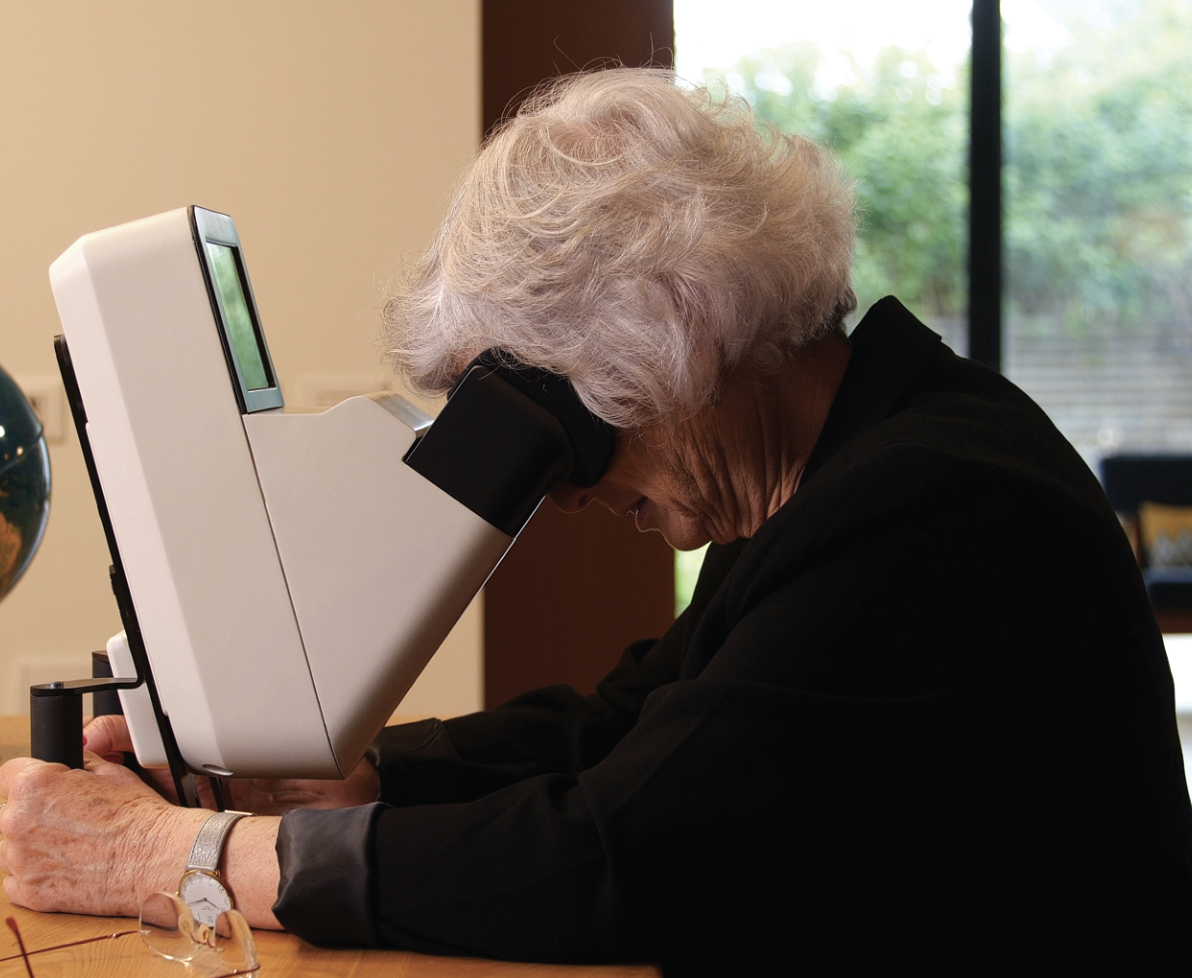 |
| A patient self-images with the Home OCT (Notal Vision) home-based optical coherence tomography device. |
Ophthalmology wasn’t ready
At its core, the CUT study showed that ophthalmology wasn’t prepared for real-time telemedicine. Although asynchronous visits have long been standard in teleophthalmology with physicians classically using the store-and-forward method—interpreting scans remotely or after the patient has left—a synchronous visit is logistically more complicated.
A patient would need to have access to some type of remote slit lamp to perform live ophthalmoscopy—technology that’s more advanced than that which is currently available to diagnose a condition in real time. Clearly, technological advances are needed to allow ophthalmologists to complete a more comprehensive synchronous ophthalmic exam/evaluation.
AI and telemedicine in DR
Proper use of telemedicine could increase the pool of people being seen by both increasing appointment efficiency and providing greater access to those not currently being monitored for retinopathy.
Some primary-care clinics have started experimenting with telehealth for DR management; first, with a remote patient evaluation, and then, second, referring the patient if symptoms are detected.
A Toronto-based, urban pilot screening program used a decision tree model to sort 566 patients into remote-manageable and in-person-visit-needed categories.5 In this program, a patient identified as at-risk during a primary-care visit would be referred to the tele-retina program. The patient would then follow-up with a mobile ophthalmic clinic where a technician would dilate, image, take intraocular pressure and check visual acuity. The fundus image would be uploaded to a server, a retina specialist would evaluate the image, and the patient would be referred for an in-person, ophthalmic visit if positive for DR.
This triaging protocol helped prioritize specialists’ time and sort DR patients efficiently. The study found that, versus a control group of in-person ophthalmic visits, tele-retina visits cost less ($109.29 vs. $315.22 Canadian per case diagnosed) and diagnosed more patients with DR (143 diagnosed in tele-retina visits vs. 79 diagnosed in standard-of-care in-person visits to initially identify DR).5
Arguably, the most exciting frontier in the evolution of teleophthalmology is the use of artificial intelligence in image analysis, the autonomous evaluation of fundus images for DR. Researchers imagine the final form of AI to be seamless imaging, analysis and generation of an ophthalmic referral if positive.
Improving AI evaluation incrementally
While we’re not there yet, researchers are incrementally improving AI evaluation. In 2008, Michael Abramoff, MD, PhD, and colleagues built the first autonomous classifier: ARIAS (automated retinal image analysis system).6
In 2013, the refined model classified referable DR with a 96.8-percent sensitivity and a 59.4-percent specificity.6 In the context of AI, sensitivity is the probability that given an image is positive for DR, the image will be marked as positive. Specificity is the probability that given an image is negative for DR, the image will be marked as negative. As of 2023, the ARIAS system, with further improvements, is used in the Scottish DR grading system as a first line of detection.6
Fundamentally, an AI-based identification of disease must perform similarly to clinicians in one or more areas. A 2023 Stanford study used the LumineticsCore reading system, formerly known as IDx-DR, one of the first Food and Drug Administration-approved autonomous, AI DR classifiers, and compared its interpretations with a team of physician readers on a macula-centered and an optic nerve-centered fundus photograph.7
 |
In the detection of more-than-mild DR, researchers found LumineticsCore’s sensitivity and specificity to be 95.5 and 60.3 percent, respectively. Remote specialists showed a sensitivity and specificity of 69.5 and 96.9 percent.7 The study supported the use of AI in triaging DR and classifying patients for first detection. With DR prevalence predicted to increase by more than 50 percent globally in the next 30 years, AI could be the medical community’s answer for quick and efficient triaging.8
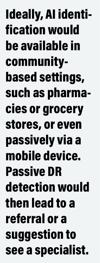
The fundamental value add is that, via an autonomous AI modality, we can produce an earlier diagnosis of DR in patients who otherwise may not have access to timely evaluation. Ideally, AI identification would be available in community-based settings, such as pharmacies or grocery stores, or even passively via a mobile device (which we look at numerous times a day with both eyes), just like pedometers. Passive DR detection would then lead to a referral or a suggestion to see a retina specialist.
Telemedicine in AMD management
AMD is amenable to observation and treatment when needed. Conversion to wet AMD is frequent enough that dry AMD patients should be monitored, and higher-risk patients or those with active exudation need to be properly identified to receive treatment in a timely manner.9 Using AI and teleophthalmology, monitoring between in-person visits could be one way to better identify patients with recurrent exudation before symptomatic vision loss.
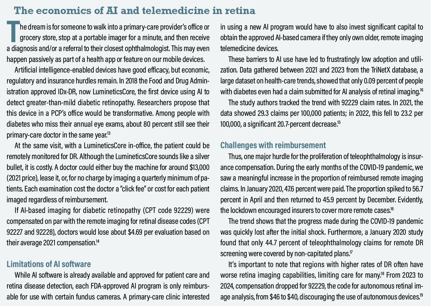
One recent remote monitoring study used the Notal Vision OCT device (Figure above), a portable device that estimates the volume of fluid in the macula.11 Forty wet AMD patients, each producing an average of six scans a week per eye, were both managed normally by an ophthalmologist and used the Notal OCT Analyzer (NOA) machine almost daily.
Of 35 in-office scans that came up as positive for fluid, the NOA found fluid in 31, an 89 percent accuracy rate. For 14 in-office scans that were negative for fluid, the NOA agreed on 10, a rate of 71 percent. The study authors described this as acceptable accuracy and sufficient for remote monitoring of stable patients who take frequent home scans.10
 |
Telemedicine and treat-and-extend
At-home testing seems particularly useful in treat-and-extend models where physicians dose patients on longer and longer intervals. Currently, ophthalmologists will extend patients by an extra one to two weeks. With at-home testing, patients could be managed with a more powerful data-driven approach based on the exact timing of detected exudation, ensuring a balance between adequate treatment and the burden of treatment.
With new, high-cost treatments such as aflibercept 8 mg (Eylea HD, Regeneron Pharmaceuticals) and faricimab (Vabysmo, Genentech/Roche) that promise extended dosing intervals, at-home testing could work as a safeguard during longer gaps between treatments.11
Many patients find it difficult financially and logistically to come in monthly. Longer treatment intervals with at-home testing would both provide insurance against silent, sudden bleeds and/or exudation when an interval proves too long, allowing patients to come in only when necessary. An OCT with at-home monitoring would be a significant improvement over the current treat-and-extend model.
AI and deep-learning models have also been proven to be accurate and applicable in AMD detection and prediction. One deep-learning model using OCT fovea images correlated a known incipient AMD biomarker with a new, unknown biomarker, highlighting the pre-wet AMD feature to researchers: hyporeflective outer retinal bands.12
Another particularly exciting model using a deep-learning algorithm predicted, with acceptable error, the conversion of nonexudative AMD to exudative AMD in both the short (three months) and long term (21 months).12
Bottom line
David Glasser, MD, at the Wilmer Eye Institute at Johns Hopkins University in Baltimore, has made the point that the more equipment-heavy a subspeciality is, the less amenable its exam will be to remote treatment.20 Unfortunately, this gap will only grow with the current trend of dwindling reimbursement.
The real tragedy, of course, is that the technology is already here, but the economics of reimbursement, regulatory hurdles, workflow and infrastructure hold back its utilization—real challenges standing in the way of AI and teleophthalmology to realize their full potential. If physicians and advocates can move the needle and strengthen teleophthalmology’s current tools, countless patients will benefit.
REFERENCES
1. Land MR, Patel PA, Bui T, et al. Examining the role of telemedicine in diabetic retinopathy. J Clin Med. 2023;12:3537.
2. Ricur G, Reyes J, Alfonso E, Marino RG. Surfing the COVID-19 tsunami with teleophthalmology: The advent of new models of eye care. Curr Ophthalmol Rep. 2023;11:1-12.
3. Aguwa UT, Aguwa CJ, Repka M, et al. Teleophthalmology in the era of COVID-19: Characteristics of early adopters at a large academic institution. Telemed J E-Health Off J Am Telemed Assoc. 2021;27:739-746.
4. Portney DS, Zhu Z, Chen EM, et al. COVID-19 and use of teleophthalmology (CUT Group): Trends and diagnoses. Ophthalmology. 2021;128:1483-1485.
5. Stanimirovic A, Francis T, Shahid N, et al. Tele-retina screening of diabetic retinopathy among at-risk populations: An economic analysis. Can J Ophthalmol. 2020;55:8-13.
6. Than J, Sim PY, Muttuvelu D, et al. Teleophthalmology and retina: A review of current tools, pathways and services. Int J Retina Vitr. 2023;9:76.
7. Dow ER, Khan NC, Chen KM, et al. Artificial intelligence-human hybrid workflow enhances teleophthalmology for the detection of diabetic retinopathy. Ophthalmol Sci. 2023;3:100330.
8. Tan TE, Wong TY. Diabetic retinopathy: Looking forward to 2030. Front Endocrinol. 2022;13:1077669.
9. Parikh R, Avery RL, Saroj N, Thompson D, Freund KB. Incidence of new choroidal neovascularization in fellow eyes of patients with age-related macular degeneration treated with intravitreal aflibercept or ranibizumab. JAMA Ophthalmol. 2019;137:914-920.
10. Blinder KJ, Calhoun C, Maguire MG, et al. Home OCT Imaging for newly diagnosed neovascular age-related macular degeneration: A feasibility study. Ophthalmol Retina. 2024;8:376-387.
11. Rowe LW, Ciulla TA. Long-acting delivery and therapies for neovascular age-related macular degeneration. Expert Opin Biol Ther. 2024;24:799-814.
12. Crincoli E, Sacconi R, Querques L, Querques G. Artificial intelligence in age-related macular degeneration: State of the art and recent updates. BMC Ophthalmol. 2024;24:121.
13. Gibson DM. Estimates of the percentage of US adults with diabetes who could be screened for diabetic retinopathy in primary care settings. JAMA Ophthalmol. 2019;137:440.
14. Chen EM, Chen D, Chilakamarri P, Lopez R, Parikh R. Economic challenges of artificial intelligence adoption for diabetic retinopathy. Ophthalmology. 2021;128:475-477.
15. Parikh R. Utilization of Autonomous AI-Based Detection of Diabetic Retinopathy Using Retinal Fundus Photography in the United States. Presented at: 2024 The Retina Society Meeting; September 12, 2024; Lisbon, Portgual.
16. Lee SC, Alber S, Lieng MK, Emami-Naeini P, Yiu G. Teleophthalmology using remote retinal imaging during the COVID-19 pandemic. Telemed J E-Health Off J Am Telemed Assoc. 2023;29:81-86.
17. Ellis MP, Bacorn C, Luu KY, et al.. Cost analysis of teleophthalmology screening for diabetic retinopathy using teleophthalmology billing codes. Ophthal Surg Lasers Imaging Retina 2020;51:S26–S34.
18. Drench DD, Behrens JJ, Jackson KL, et al. Payment Reform Needed to Address Health Disparities of Undiagnosed Diabetic Retinopathy in the City of Chicago. Ophthalmol Ther. 2017;6:123-131. doi:10.1007/s40123-016-0072-4
19. CMS-1784-F. CY2024 Payment Policies Under the Physician Fee Schedule. Federal Register November 16, 2023. https://www.federalregister.gov/documents/2023/11/16/2023-24184/medicare-and-medicaid-programs-cy-2024-payment-policies-under-the-physician-fee-schedule-and-other.
20. Glasser DB. Is there a future for telehealth in ophthalmology? JAMA Ophthalmol. 2023;141:61.